 | Info
Sheets |
| | | | | | | | | | | | | | | | | | | | | | | | |
 | Out-
side |
| | | | |
|
| | | | | |  | Searchterm 'Surface Coil' was also found in the following services: | | | | |
|  |  |
| |
|
( SNR or S/N) The signal to noise ratio is used in MRI to describe the relative contributions to a detected signal of the true signal and random superimposed signals ('background noise') - a criterion for image quality.
One common method to increase the SNR is to average several measurements of the signal, on the expectation that random contributions will tend to cancel out. The SNR can also be improved by sampling larger volumes (increasing the field of view and slice thickness with a corresponding loss of spatial resolution) or, within limits, by increasing the strength of the magnetic field used. Surface coils can also be used to improve local signal intensity. The SNR will depend, in part, on the electrical properties of the sample or patient being studied.
The SNR increases in proportion to voxel volume (1/resolution), the square root of the number of acquisitions ( NEX), and the square root of the number of scans ( phase encodings). SNR decreases with the field of view squared (FOV2) and wider bandwidths. See also Signal Intensity and Spin Density.
Measuring SNR:
Record the mean value of a small ROI placed in the most homogeneous area of tissue with high signal intensity (e.g. white matter in thalamus). Calculate the standard deviation for the largest possible ROI placed outside the object in the image background (avoid ghosting/aliasing or eye movement artifact regions).
The SNR is then:
Mean Signal/Standard Deviation of Background Noise | | | | | | | | | | | | |  Further Reading: Further Reading: | | Basics:
|
|
News & More:
| |
| |
| | | | | |
| |
|
From Toshiba America Medical Systems Inc.;
the Ultra™ system was developed to help healthcare providers be more competitive by delivering greater patient comfort and a broad range of clinical capabilities, says Anita Bowler, product manager, MRI Business Unit, Toshiba America Medical Systems Inc With its unique, powerful gradient technology, the Ultra™ performs advanced clinical studies and consistently provides high-resolution images that are typically associated with high field MRI systems. At the same time, the Ultra™ offers a truly open feeling that makes patients more relaxed, especially those with claustrophobic tendencies.
Device Information and Specification CLINICAL APPLICATION Whole Body Quadrature, solenoid and multi-channel configurations SE, FE, IR, FastSE, FastIR, FastFLAIR, Fast STIR, FastFE, FASE, Hybrid EPI, Multi Shot EPI, Single shot EPI diffusion, True SSFP, SuperFASE; Angiography: 2D(gate/non-gate)/3D TOF, SORS-STC, Black Blood MRA POWER REQUIREMENTS 380/400/415/440/480 V COOLING SYSTEM TYPE Cryogenless | | | | | |
| | | | | |
| |
|
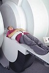
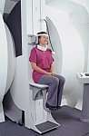
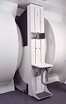
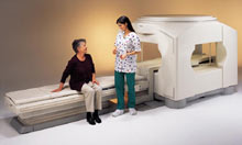
From FONAR Corporation;
in October of 2004, the company changed the product name of the Stand-Up™ MRI to the Upright™ MRI. The Indomitable™, Upright™ MRI is the only open MRI in the world that can perform positional MRI (pMRI), i.e. the Upright MRI™ scans patients in upright, weight-bearing positions, in addition to the conventional lie-down positions. The Upright™ MRI is the only device that can scan patients in the position their symptoms occur, in their position of pain. In early clinical reports independently confirm the effectiveness and potential of positional MRI. In October 2000, Fonar received permission to market the Indomitable™ from the FDA.
Device Information and Specification CLINICAL APPLICATION Whole body -
weight-bearing MRI -
position imaging (flexion, extension, bending, standing, sitting and recumbent scanning)
CONFIGURATION Front-open and Top-open MRIPOWER REQUIREMENTS 380/400/415/440/480 V COOLING SYSTEM TYPE Water, closed-loop | | | |
• View the DATABASE results for 'Upright™ MRI' (5).
| | |
• View the NEWS results for 'Upright™ MRI' (9).
| | | | | | Further Reading: | Basics:
|
|
News & More:
| |
| |
| | | Searchterm 'Surface Coil' was also found in the following services: | | | | |
| | |
| |
|
From Toshiba America Medical Systems Inc.;
VISART™ series is a 1.5 Tesla superconducting MRI system that has been designed to meet the expanding role of MRI in today's clinical environment. The system utilizes innovative technologies such as digital RF, high speed actively shielded gradients and optimized RF coils, which support a wide range of MRI developments. The Visart, an early type of Toshiba Medical Systems Inc., can be retrofitted or upgraded to the Excelart configuration.
Device Information and Specification CLINICAL APPLICATION Whole body Quadrature, solenoid and multi-channel configurations SE, FE, IR, FastSE, FastIR, FastFLAIR, Fast STIR, FastFE, FASE, Hybrid EPI, Multi Shot EPI; Angiography: 2D(gate/non-gate)/3D TOF, SORS-STC IMAGING MODES Single, multislice, volume study POWER REQUIREMENTS 380/400/415/440/480 V COOLING SYSTEM TYPE Closed-loop water-cooled | | | |
• View the DATABASE results for 'VISART™' (2).
| | | | | | Further Reading: | News & More:
|
|
| |
| | | | | |
| |
|
From Toshiba America Medical Systems Inc.;
With its high-field strength, the Vantage™ delivers the clinical capabilities and image quality expected by cardiologists, while simultaneously offering patients a more comfortable and non-invasive option, said Dane Peshe, director, MRI Business Unit, Toshiba America Medical Systems. Vantage™ supports a full complement of cardiovascular imaging studies, ranging from stroke evaluation to peripheral vascular imaging. Additionally, the ultra short bore design offers patients a greater feeling of openness that reduces claustrophobic sensations, while Toshiba's exclusive, patented Pianissimo™ technology reduces scan noise by as much as 90 percent for a more pleasant experience.'
Device Information and Specification CLINICAL APPLICATION Whole body CONFIGURATION Ultra short bore SE, FE, IR, FastSE, FastIR, FastFLAIR, Fast STIR, FastFE, FASE, EPI, SuperFASE; Angiography: 2D(gate/non-gate)/3D TOF, SORS-STC IMAGING MODES Single, multislice, volume study 32-1024, phase;; 64-1024, freq. POWER REQUIREMENTS 380/400/415/440/480 V COOLING SYSTEM TYPE Closed-loop water-cooled Liquid helium: approx. less than 0.05 L/hr Passive, active, auto-active | | | |
• View the DATABASE results for 'Vantage™' (2).
| | | | | | Further Reading: | Basics:
|
|
| |
| | | | |
| | | |
|
| |
 | Look
Ups |
| |


